Tissue Microarray Construction

New TMA Construction | Fees | Forms |TMA array maps | Links | Contact

Overview

a picture of TMAs being constructed

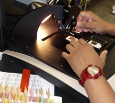
Our facility is used for the construction of tissue microarrays (TMAs). TMAs are a powerful new technology for high throughput analysis of biomarker expression in a large number of tissue samples. These samples range from human tissue specimens with clinical follow-up, animal tissues, to cell lines.

The TMA Lab has constructed 900 TMAs for 75 investigators containing 145,000 tissue cores from tumors from the prostate, breast, pancreas, gall bladder, cervix, ovary, brain, liver, urinary bladder, salivary glands, esophagus, head and neck, thyroid, adrenal, xenografts, autopsy, colon, skin, testis, kidney, lung and various cell lines. Users have published >150 manuscripts involving JHU TMAs.

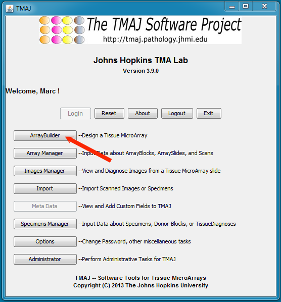
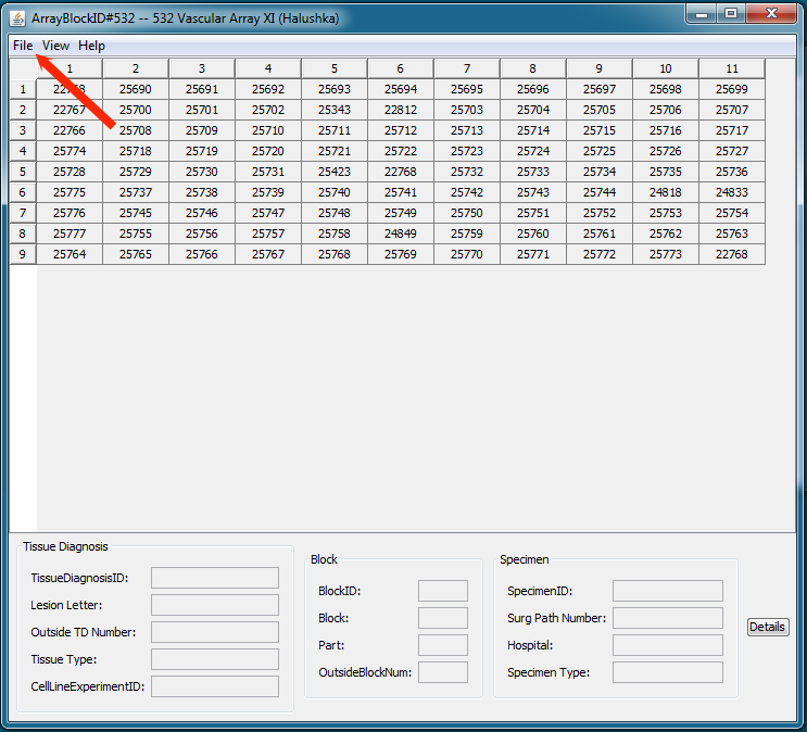
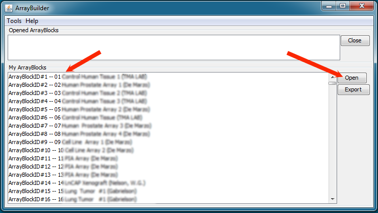
Instrumentation is available to automatically capture high resolution digital images of TMA spots and perform semi-automated quantitative image analysis. Users have access to a set of Open Source web-based software tools and backend database (TMAJ) that were developed to facilitate the all facets of research involving TMA technology from TMA design, data entry, image diagnosis, analysis, and publishing on the Internet.

Please email us at hfedor@jhmi.edu today to set up a free consultation to discuss your project. The TMA Core will provide free advice to investigators on array design. Investigators with an interest in TMA data handling beyond simple spreadsheets will have full access to our newly developed TMA database, TMAJ .

New Array Construction

Please read this document to learn about the steps needed to create a new TMA.

Turn around time: Depending on current demand the turn around time can vary between 5 days and 3 weeks.

Back to top

Requesting TMA Slides from an Existing TMA

If you are the PI or are designated as the PI for a particular TMA that already exists, simply fill out the requisition form including budget and IRB approval number.

If you are not the PI for the array, you may wish to contact the PI regarding using TMA slides from an existing array for your own research. We require written (email is OK) acknowledgement that it is OK for us to provide you with TMA slides from that array. Please include the number of slides.

Some PIs have agreed to share information about the TMAs they have produced and may be available for collaboration. We maintain an active list of these. Please contact Helen Fedor (hfedor@jhmi.edu)
for more information.

About Tissue MicroArrays

Tissue Microarrays (TMAs) consist of paraffin blocks where up to 500 individual tissue cores have been inserted in an arrayed fashion (1). These miniaturized collections of tissue spots result in a dramatic increase in throughput for in situ examination of gene status and gene expression from archival specimens. All available methods for examination of histological sections, including immunohistochemistry, in situ hybridization, DNA ploidy analysis, nuclear morphometry, and fluorescent in situ hybridization can be performed on sections from TMA blocks. Many clinical specimens contain limited tissues for study. TMAs can expand the number of studies that can be performed from these extremely valuable specimens. TMAs have been validated as useful tools for the evaluation of various biomarkers, especially in oncology patients.

To learn more about tissue microarrays, please visit this site at the University of Virginia which provides additional information on what TMAs are and how to make optimal TMAs.

Fees

Having constructed numerous arrays over the last several years we have arrived at a pricing structure for TMA that is commensurate with our costs. We feel that our pricing is very competitive as compared to those offered by other institutions and that we can provide significant value and expertise.

Please note that these prices are for JHU Cancer Center members. Rates for non-cancer members affliated with JHU are 10% higher. Non-JHU affiliates should send inquiry to Helen Fedor (hfedor@jhmi.edu). Prices are subject to change.
Construction Set-Up
$275.00
Cores Transfer
$2.00
Control Tissue 1.5mm
$7.00
Control Tissue 1.0mm
$3.50
Control Tissue 0.6mm
$1.50
Control Tissue slide on Chemate
$8.50
Sectioning Set-Up
$60.50
Unstained TMA Slide
$4.84
In-complete TMA Slide
$2.00

Back to top

Forms

  • We no longer offer work request forms here. All requests must go through our campus-wide iLab tool.

Back to top

TMA maps

    If you need a print out of your TMA map (indicating which core is which), please use the following protocol.

    1) Log into TMAJ. If you do not have a login to TMAJ or access to your TMA of interest, please contact Helen Fedor (hfedor@jhmi.edu).

    2) Click on ArrayBuilder Application

    3) Select and open the desired Array Block ID corresponding to your TMA.

    4) Click File, then either Excel or Export, and Alternate for the map we use to construct the array.

    5) Click Full Details instead of Alternate for a list of cores layout version.

Back to top

Links

Contact

Location:
TMA Laboratory
Johns Hopkins Pathology
411 N. Caroline Street
Basement Room #312
Baltimore, MD 21231

For inquiries, please contact:
Helen Fedor
Email: hfedor@jhmi.edu
Phone: 410-502-3358
Fax: 410-502-5158
Pager: 410-283-3419

Back to top